What do images of a fetus at 25 weeks reveal about development? Visual representations of fetal growth at this stage offer a unique window into prenatal development.
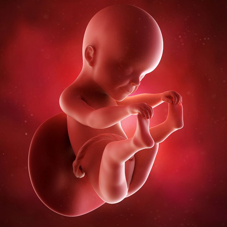
Images of a fetus at 25 weeks gestation depict a significant stage of prenatal development. These images showcase a noticeably more mature and refined structure compared to earlier stages. Characteristic features, such as well-defined limbs, discernible fingers and toes, and potentially visible facial details, are often present. Variations in image quality can occur depending on the imaging technique (e.g., ultrasound) and the specific equipment used.
Visual representations of the developing fetus at 25 weeks are crucial for medical professionals. They allow for assessment of fetal growth and well-being. Identifying any anomalies or potential concerns is significantly aided by these images. Furthermore, these images can offer reassurance to expecting parents, visually confirming the progression of development and potentially reducing anxiety. From a broader perspective, such images contribute to our understanding of the remarkable processes involved in human gestation.
Read also:Dior Goodjohn Stunning Designs Exclusive Finds
Transitioning to the following sections, we will explore the various types of imaging technologies used to capture these images, delve into the ethical considerations surrounding the use of fetal imagery, and examine how this stage of development influences subsequent stages.
25 Weeks Pregnant Fetus Images
Visualizations of fetuses at 25 weeks provide crucial information about development. These images are essential for medical professionals and expectant parents.
- Fetal development
- Growth assessment
- Medical diagnosis
- Parental reassurance
- Ultrasound technology
- Ethical considerations
- Gestational timeline
Understanding fetal development at 25 weeks involves careful analysis of images. Growth assessment relies on these images to monitor progress against expected norms. Medical diagnoses may identify abnormalities, impacting subsequent care. Visual confirmation can offer reassurance, mitigating parental anxiety. Ultrasound technology generates these images, requiring careful interpretation. Ethical considerations accompany the use of fetal imagery. The gestational timeline is crucial for interpreting the images and understanding development stages. For example, well-defined limbs and facial features observed through an ultrasound image at 25 weeks confirm expected progression. These details collectively underscore the importance of fetal images in prenatal care and understanding fetal development within a specific gestational window.
1. Fetal Development
Fetal development at 25 weeks is a critical stage characterized by significant maturation. Visual representations, such as those obtained through imaging, are vital for understanding and assessing this progress. Images at this juncture provide concrete evidence of developmental milestones, informing medical professionals and expectant parents.
- Organ System Maturation
At 25 weeks, organs continue to develop and refine. Images reveal progressing complexity in organ structure and function. For example, the lungs, though not fully developed, begin to produce surfactant, a crucial substance for respiration. The maturation of the nervous system is also evident in images, as the brain becomes more defined and intricate. These observable developmental changes provide valuable data for assessing the overall health of the fetus.
- Growth and Size
Images at 25 weeks demonstrate characteristic growth and size parameters. Accurate measurements are critical for evaluating if the fetus is developing at a normal rate compared to gestational norms. Significant disparities in growth can indicate potential complications. These measurements from images support diagnostic decision-making.
Read also:
- Natural Oleato Benefits Uses
- Physical Characteristics
At 25 weeks, crucial physical characteristics are observable. Images demonstrate the presence of well-defined limbs and digits. Facial features start to become more recognizable, with refined features. Consistent visual data helps evaluate overall fetal well-being and serves as a point of reference compared to expected development timelines.
- Neurological Development
Although fully developed at later stages, neurological development is evident by 25 weeks. Images can depict brain structure and activity. While subtle, these indicators assist in assessing neurological function. Subtle changes, indicative of developing neurological pathways, are crucial in understanding fetal potential.
In summary, images of a fetus at 25 weeks provide a crucial snapshot of developmental progress. These images allow for crucial assessments across multiple facets of development, from organ maturation to growth patterns, physical characteristics, and neurological progression. This data is vital for making informed decisions regarding the ongoing health of the fetus.
2. Growth Assessment
Accurate assessment of fetal growth at 25 weeks is critical. Images obtained through various technologies provide crucial data for evaluating fetal development against expected norms. This analysis informs medical decisions and guides subsequent care strategies.
- Measurement Techniques
Precise measurements are derived from imaging. Ultrasound, in particular, offers a non-invasive method for determining fetal size and biometric parameters. These measurements, including head circumference, abdominal circumference, and femur length, are crucial for comparing to established growth charts. Variations from these norms can indicate potential developmental issues, necessitating further investigation. The accuracy of these measurements significantly influences subsequent interpretations and clinical interventions.
- Growth Charts and Norms
Growth charts serve as benchmarks for healthy fetal development. These charts provide age-specific thresholds for appropriate fetal size. Images allow comparisons to these charts to assess if a fetus is growing within the expected range for its gestational age. Discrepancies can signal the need for further medical assessment, identifying potentially underlying factors like placental insufficiency or genetic conditions.
- Identification of Potential Issues
Growth assessment, aided by images, assists in the early identification of potential problems. Substantial deviations from typical growth patterns can be indicators of underlying medical issues affecting fetal well-being. Images facilitate comparison of current development to expected norms, enabling prompt medical intervention if required. This early detection is crucial in maximizing positive outcomes.
- Long-Term Implications
Assessment of growth at 25 weeks has implications for subsequent fetal development. Early identification of growth issues may necessitate interventions to optimize fetal growth and well-being. Subsequent developmental milestones can be affected, and proper intervention can mitigate negative consequences. Appropriate interventions depend directly on the accurate interpretation of images in growth assessment protocols.
In conclusion, growth assessment utilizing images of fetuses at 25 weeks is integral to prenatal care. Precise measurements, comparisons to growth charts, and the early detection of growth deviations enable proactive medical interventions. This process ultimately aims to ensure optimal fetal development and overall well-being.
3. Medical Diagnosis
Accurate medical diagnosis relies heavily on precise information derived from various sources, including images of a fetus at 25 weeks gestation. The presence and clarity of anatomical structures, as visualized in these images, offer valuable clues for evaluating fetal health and identifying potential abnormalities.
- Identifying Structural Anomalies
Visual analysis of images can reveal structural abnormalities that might not be apparent otherwise. These images, frequently obtained through ultrasound, allow for the assessment of organs and their proper placement. Difficulties in development, such as heart malformations, neural tube defects, or limb abnormalities, may be detected. The clarity and detail present in 25-week fetal images aid in identifying subtle but critical anomalies. Early diagnosis allows for prompt intervention and management strategies.
- Evaluating Fetal Growth and Development
Fetal growth assessment is a crucial component of medical diagnosis. Images provide a visual record of the fetus size, shape, and development at 25 weeks. Comparing these measurements against standard growth charts helps identify deviations that could signal underlying conditions such as chromosomal abnormalities or placental insufficiency. A proper diagnosis informed by this data helps in providing personalized medical care.
- Assessing Fetal Well-being
Images offer an assessment of the overall well-being of the fetus. Signs of distress, such as abnormal amniotic fluid levels or decreased fetal movement, may be detected visually. These observations, combined with other clinical data, assist in assessing the overall health status of the fetus and guide decisions regarding further monitoring or intervention.
- Monitoring Complications
For pregnancies with known complications or risk factors, regular imaging plays a critical role in monitoring their progression and response to treatment. Images at 25 weeks can provide crucial data to evaluate the impact of factors such as gestational diabetes or hypertension on fetal development and health. Continued monitoring and analysis of successive images aid in making well-informed decisions in managing complex pregnancies.
In conclusion, images of a fetus at 25 weeks are integral to medical diagnosis. The visual data allows for the identification of structural anomalies, assessment of growth and development, evaluation of fetal well-being, and monitoring of complications. These diagnostic insights directly influence management strategies, improving outcomes for both the fetus and the expectant parent. The information derived from these images informs clinical decisions, offering valuable tools for effective prenatal care.
4. Parental Reassurance
Images of a fetus at 25 weeks gestation play a significant role in providing reassurance to expecting parents. Visual confirmation of fetal development, health, and progress can alleviate anxieties and promote a sense of confidence during this critical stage of pregnancy. The clarity and detail in these images, often depicting recognizable features, contribute to a more tangible connection between parents and their developing child.
- Visual Confirmation of Progress
Images offer a tangible representation of fetal development, illustrating the stages of growth and maturation. Seeing the progress visually can offer substantial reassurance, counteracting the natural anxieties associated with pregnancy. The discernible features in the images, such as developed limbs, facial characteristics, and organ structure, provide a strong visual testament to the health and well-being of the fetus.
- Addressing Concerns and Anxiety
Visual confirmation of fetal development can address concerns and anxieties related to the pregnancy. Potential anxieties regarding fetal growth, health issues, or genetic predispositions can be mitigated by the clear and comprehensive data presented in these images. The reassurance derived from these images contributes to a more positive emotional environment for expecting parents, enhancing their confidence and preparedness for the challenges and joys of parenthood.
- Enhanced Parental Connection
Images facilitate a deeper emotional connection between parents and their developing child. The visible stages of development and the clarity of the fetus's form can create a more personal and meaningful experience for parents. Seeing the developing features can nurture anticipation and emotional bonding, fostering a stronger relationship between parent and child even before birth.
- Facilitating Informed Decisions
In instances where concerns arise or abnormal findings are observed, these images contribute to informed decision-making. The clarity and detail enable healthcare professionals to communicate findings effectively to parents, fostering open communication and collaboration. The images allow for a deeper comprehension of the situation and enable parents to make well-informed choices based on the observable data.
In conclusion, images of a 25-week fetus are crucial for parental reassurance, offering a visual confirmation of development, addressing anxiety, fostering connection, and enabling informed choices. These images are not merely diagnostic tools but vital components of the emotional support system for expecting parents.
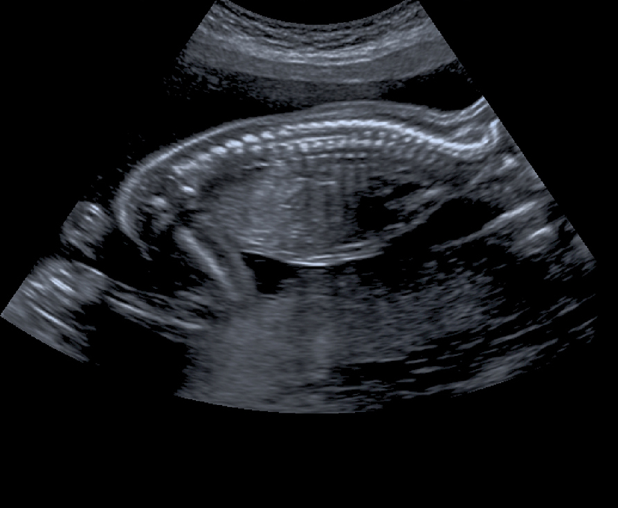
5. Ultrasound Technology
Ultrasound technology is fundamental to acquiring images of a 25-week-pregnant fetus. This non-invasive technique employs high-frequency sound waves to create detailed images of the developing fetus. The process allows for visualization of internal structures, facilitating assessment of fetal growth, development, and overall well-being. Sophisticated ultrasound machines, with their advanced image processing capabilities, contribute significantly to the quality and detail of the resulting images. The precise timing of the 25-week ultrasound is crucial for capturing crucial developmental milestones, enabling healthcare professionals to evaluate the fetus's progress against established norms.
The utility of ultrasound extends beyond mere visualization. Accurate measurements derived from these images are crucial for assessing fetal growth parameters, such as head circumference, abdominal circumference, and femur length. Deviations from established norms can signal potential complications, necessitating further investigation and interventions. Moreover, ultrasound aids in the identification of structural abnormalities. Early detection of conditions like heart defects or neural tube defects is critical for implementing appropriate management strategies and impacting the long-term prognosis. Examples include cases where early ultrasound detection of a neural tube defect enables targeted interventions, potentially improving outcomes. The technology's ability to visualize the developing organs and structures contributes to the accuracy of such diagnoses. The non-invasive nature of ultrasound is paramount for repeated assessments throughout pregnancy, facilitating ongoing monitoring of fetal development and health without causing harm.
In conclusion, ultrasound technology is indispensable in producing images of a 25-week-pregnant fetus. Its ability to provide detailed anatomical images, coupled with accurate measurements and the early detection of potential anomalies, is crucial for effective prenatal care. The non-invasive nature of this technology allows for repeated assessments, thereby contributing significantly to the successful monitoring and management of pregnancies. While ultrasound technology is not without limitations, its widespread use and advancement in technology continue to enhance the precision and reliability of prenatal assessments. The information gleaned from these images shapes critical decisions regarding fetal health and contributes to the comprehensive care of the expectant mother and the developing fetus.
6. Ethical Considerations
Ethical considerations surrounding images of a 25-week-pregnant fetus are multifaceted and arise from the inherent value attributed to the developing human life. The ability to visualize a fetus at this stage raises complex questions regarding the use of such imagery, particularly concerning the balance between scientific advancement and the potential for emotional impact on individuals involved. Decisions about the use and dissemination of these images must be made with careful consideration for the implications for both the pregnant person and the fetus. The visual nature of the images makes them particularly potent in shaping perceptions and influencing decisions regarding the course of a pregnancy.
Practical application of ethical frameworks in the context of 25-week fetal images requires sensitivity and transparency. Potential dilemmas include the use of fetal images for diagnostic purposes, the potential for misinterpretation of images and their implications for emotional distress, and the right of the pregnant individual to access or refuse access to such images. Informed consent is crucial. The pregnant individual should be fully informed about the potential implications of diagnostic imaging, including the potential for distressing outcomes in cases of unforeseen anomalies, and must have the autonomy to choose whether to pursue such procedures. Cases involving legal disputes surrounding fetal imaging, especially regarding the right to an abortion or the rights of parental figures, highlight the urgent need for clear ethical guidelines. A thorough understanding of applicable legal and ethical standards is imperative to navigate these complex situations. For instance, if an image reveals a condition that impacts the potential choices regarding the pregnancy, the communication of this information must adhere to ethical considerations regarding patient autonomy and emotional well-being.
In conclusion, ethical considerations are paramount when examining images of a 25-week-pregnant fetus. The potential for profound emotional impact on both the pregnant individual and their support system demands that healthcare providers and imaging professionals approach these images with sensitivity and adherence to robust ethical protocols. Clear guidelines, informed consent processes, and a commitment to patient autonomy are essential components of responsible fetal imaging practices. The integration of ethical considerations into the framework of medical imaging for fetuses at this stage safeguards the well-being of all parties involved, ensuring that technological advancements are utilized responsibly and respectfully.
7. Gestational Timeline
The gestational timeline provides a framework for understanding fetal development. Precisely pinpointing the stage of gestation, like 25 weeks, is crucial for interpreting images. The timeline establishes expectations for the size, structure, and functionality of the fetus. Variations from the expected timeline can signal potential issues, requiring further medical assessment.
- Developmental Milestones
The gestational timeline outlines expected developmental milestones at each week. At 25 weeks, specific markers of development, such as the degree of lung maturation, central nervous system function, and limb development, are anticipated. Images at 25 weeks should reflect these expected developmental characteristics. Differences between observed development in images and the expected timeline may indicate a need for further medical investigation.
- Growth and Size Standards
A key component of the timeline involves fetal growth patterns. Images taken at 25 weeks allow for a comparison of fetal size and proportions with established growth standards. These standards are based on large populations of healthy pregnancies and provide benchmarks for evaluating whether fetal development aligns with expectations. Deviations from expected growth patterns observed in images can indicate possible underlying health conditions.
- Organ System Maturation
The timeline encompasses the maturation of various organ systems. At 25 weeks, specific organ systems are expected to be at particular stages of development. Images reflecting the expected levels of development can help ensure proper fetal health and growth. Any significant deviation from this timeline as observed in the images would warrant further medical attention.
- Interpretation of Imaging Results
The gestational timeline is essential for correctly interpreting the findings from 25-week fetal images. Anomalies, if detected, must be placed within the context of the expected developmental stage. Knowing the gestational age allows medical professionals to accurately contextualize observed characteristics in the images. Images must be critically evaluated in light of the precise gestational age for correct diagnosis.
In essence, the gestational timeline provides a crucial framework for interpreting images of a 25-week-pregnant fetus. The images offer a snapshot of development, while the timeline contextualizes this snapshot. This connection allows for a comprehensive understanding of the fetus's well-being and guides appropriate medical interventions when deviations are identified. A strong correlation between the observed characteristics in the images and the expected developmental milestones at 25 weeks is vital for accurate assessment and treatment planning. Deviances, as revealed in the images, require consideration against the gestational timeline to determine the appropriate action.
Frequently Asked Questions about 25-Week Pregnant Fetus Images
This section addresses common inquiries regarding images of fetuses at 25 weeks gestation. The information provided is for educational purposes only and should not be substituted for professional medical advice. Consult a healthcare provider for personalized guidance.
Question 1: What do 25-week fetal images typically depict?
Images of a fetus at 25 weeks gestation generally reveal significant developmental progress. Characteristic features, such as well-defined limbs, discernible fingers and toes, and increasingly detailed facial features, are often present. The visibility of these structures aids in assessing overall fetal health and growth. The quality and clarity of the images can vary based on the imaging technique (e.g., ultrasound) and equipment used.
Question 2: How are these images used in medical practice?
Images at 25 weeks are crucial for evaluating fetal growth and well-being. Measurements derived from these images, such as head circumference and abdominal circumference, are compared to established norms. Deviations from these norms may indicate potential developmental or health issues requiring further investigation and care. The visualization of anatomical structures in the images aids in identifying potential structural abnormalities. Images also serve as a benchmark for subsequent assessments, tracking progress and monitoring the response to treatment when necessary.
Question 3: What are the ethical considerations associated with these images?
Ethical considerations surround the use of fetal images. Informed consent is paramount, ensuring the pregnant person understands the implications of imaging, including the potential emotional impact of the results, both positive and potentially negative. Images should be used responsibly and ethically, prioritizing the well-being of the pregnant person and the developing fetus. Healthcare providers must carefully consider the potential for distress and handle situations with sensitivity and respect for individual autonomy.
Question 4: Are there limitations to the information provided by these images?
While informative, 25-week fetal images are not exhaustive. These images primarily visualize physical structures and characteristics, providing limited insight into the functional capacity of the fetus. Further assessments, including additional imaging or clinical evaluations, may be necessary to fully evaluate fetal well-being and inform subsequent care strategies. Images should be viewed as part of a comprehensive assessment, not as standalone proof of fetal health.
Question 5: How does the gestational timeline influence image interpretation?
The gestational timeline provides crucial context for interpreting fetal images at 25 weeks. Developmental milestones, growth standards, and anticipated organ maturation are all established within this framework. Images are evaluated against these standards to determine if fetal development aligns with expectations. Deviations from the expected timeline may necessitate further evaluation to understand the cause and implications for the ongoing pregnancy. The precise gestational age influences the interpretation of all observed features in the images.
Understanding the information presented in 25-week fetal images is essential for both medical professionals and expectant parents. Accurate interpretation and application of this data contribute to effective prenatal care and support positive outcomes.
The following sections will explore the diverse methods of fetal imaging, along with their relative benefits and drawbacks.
Conclusion
Images of a 25-week-pregnant fetus offer a critical window into prenatal development. These images, often obtained through ultrasound, facilitate the assessment of fetal growth, structure, and overall well-being. Accurate measurements derived from these images are compared to established norms to identify potential deviations from expected development. The visualization of anatomical structures allows for the detection of structural abnormalities, facilitating early intervention and management strategies. Visual confirmation of developmental milestones can provide reassurance to expecting parents. However, interpretation necessitates careful consideration of the gestational timeline, ensuring accurate context for observed characteristics. Ethical considerations regarding the use and implications of these images are paramount. The combined insights from these images contribute significantly to the comprehensive care of the developing fetus and the expectant parent.
Fetal imaging at 25 weeks serves as a vital component of prenatal care, providing a means for early identification of potential issues. The ability to visualize structural development, assess growth parameters, and monitor overall fetal health allows for informed decision-making throughout the remainder of the pregnancy. Future research should continue to refine imaging technologies and interpretation protocols to optimize the accuracy and safety of these assessments. Furthermore, ongoing dialogue regarding ethical considerations surrounding fetal imaging remains crucial for responsible practice in this sensitive area. The utilization of these images for proactive care fosters the potential for improved outcomes for both the developing fetus and the expectant parent.